
Students in the Anderson University Pathologists’ Assistant program brought the microscopic world into a macroscopic view, engaging in a creative project to display one of four main tissue types creatively. This unique merging of art and science is one of many examples of Anderson University’s pillar of Great Academics.
The students spent three weeks before spring break learning about the microscopic characteristics, functions and locations of the four main tissue types found within the human body—connective, nervous, muscle and epithelium.
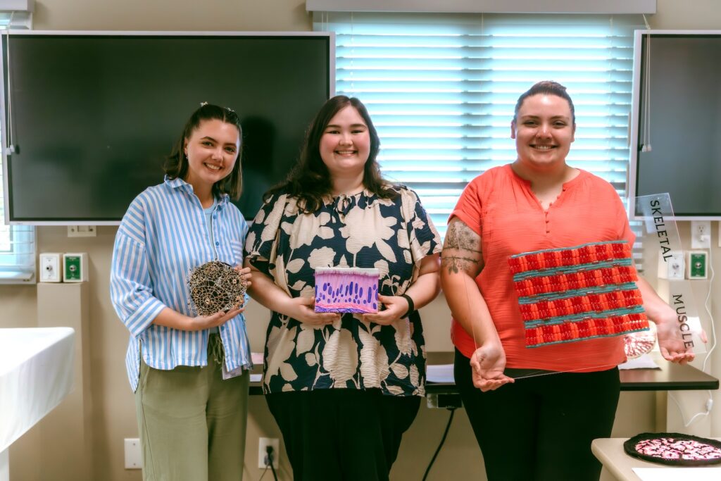
Three students whose projects scored high talked about their projects.
Colleen Heuser was drawn to display ciliated pseudostratified columnar epithelium—a tissue found in the upper respiratory tract that traps inhaled debris. Heuser’s painting on canvas represents the tissue after H&E (Hematoxylin and Eosin) staining, a technique allowing pathologists to examine tissue and identify cell types. Goblet cells are depicted as the lighter purple, oval shapes with purple dots within them and hairlike projections known as cilia.
Heuser enjoyed tapping into her artistic side as she went to work on her project.
“It basically connects this aspect that I personally enjoy doing and it allows me to visualize and truly understand and grasp the different components that are included in this type of cell, Heuser said.
“It was a little out of the box,” admitted Aaliyah Thomas. Bouncing the idea off her father, she came up with a way of visualizing skeletal muscle tissue, the most common type of muscle tissue, which is responsible for our voluntary movements. She was working with her father, whom she considers to be very creative, imagining how she might demonstrate how the tissue appears. She came up with a“sweet” discovery. Examining a microscopic view, they concluded that the elongated cylindrical cells arranged in parallell bundles (fascicles) resemble Mike and Ike and Hot Tamale candies.
“Once I saw his vision… When I saw a way to explain it to him and teach it to him, it came all together for me,” said Thomas, who wants to enter forensic pathology.
“I like to do art in my spare time,” said Gracey (Olivia) Hefner, whose project is a sculpture depicting cortical bone tissue. Cortical bone functions as the dense, outer, hard layer which provides structural support, protection for the inner portions of the bone, and provides strength to resist various forces. “I’m a very visual person. I can remember things better if I see them physically or can recreate them physically. A lot of my assignments, I’ll draw out because it just helps me understand it better.”
“I got a Styrofoam base and painted it black so that the wire would stand out when you look at it. Then I took some wire and made different sized rings to represent the different units of cortical bone, because they’re not all the same size, they’re different depending on where you are in the body,” Hefner said. “I would connect them with a different color wire to kind of separate out the different parts, so you would be able to see them more clearly.”
The projects are on display through the remainder of the semester at the Holdredge Bearwood Center of Anderson University at 3031 N. Highway 81, Anderson, South Carolina.
The Pathologists’ Assistant program is hosted by the Anderson University School of Clinical Laboratory Sciences.